
Case Studies
Expertise Showcase
GPCRs are the membrane protein class with the team's most systematic technical accumulation and richest research experience, with long-term work on expression, purification, stability optimization, complex preparation, and Cryo-EM structural studies across GPCR subclasses.
GCGR · GLP-1R · GIPR · FFAR1 · FFAR2 · SSTR family · NMUR2
Experience with GCGR structural studies under different ligand and effector protein states.
mGlu2-4 · mGlu7 · NPY1R · NPY2R · A2AR · NTSR1 · DRD family · 5-HT receptor family
Experience with multiple mGluR subtypes and homo-/heterodimer structural studies, covering different conformations and ligand recognition mechanisms.
CCR5 · CX3CR1 · FPR2 · GPR15 · CMKLR1 · GPR1
Covering difficult structural targets such as CCR5, CX3CR1, and FPR2.
ADGRD1 · ADGRF1 · ADGRG6 · ADGRV1 · T2R14 · T2R46
Covering complex membrane protein systems beyond Class A, including bitter taste receptors.
Covering Class A, adhesion, and bitter taste receptor systems
Endocrine · Neurological · Chemokine · Immunity · Inflammation · Adhesion & Other Complex GPCRs · Metabolic
Expression · Purification · Stabilization · Cryo-EM structural analysis
Representative Research Outputs
The team's membrane protein structural research has been published in journals including Science, Nature, Nature Chemical Biology, Nature Communications, Cell Research, Science Advances, Nature Structural & Molecular Biology, and Protein & Cell.
| Research Area | Representative Targets | Representative Outputs |
|---|---|---|
| Metabolic / Endocrine GPCR | GCGR | Gs / Gi recognition and receptor activation mechanism studies |
| Neurological GPCR | mGlu2 / mGlu4 | Gi binding and receptor structure studies |
| NPY Receptors | Y1R / Y2R | Ligand recognition and receptor activation mechanisms |
| Adhesion GPCR | ADGRD1 / ADGRF1 | Tethered agonism mechanism studies |
| Chemokine Receptors | CCR5 | Ligand recognition and receptor activation mechanisms |
| Chemokine Receptors | CX3CR1 | Cholesterol-regulated receptor activation studies |
| Inflammation-related GPCR | FPR2 | Ligand recognition and neuroprotective mechanism studies |
| Reproductive / Endocrine | SSTR family | Ligand recognition and selectivity mechanism studies |
| Lipid Metabolism | FFAR1 / FFAR2 | Endogenous ligand recognition and activation mechanisms |
| Purinergic Receptors | P2Y1R / P2Y12R | Ligand recognition and signal transduction studies |
| Reproductive Endocrine | NMUR2 | Ligand binding and receptor activation mechanisms |
| Oncology / Immunity | ADGRG6 | Allosteric ligand binding mechanism studies |
| Taste Receptors | T2R14 / T2R46 | Ligand binding mode studies |
Case Study
Case 1: Cryo-EM Structure of A2AR-Gs Complex
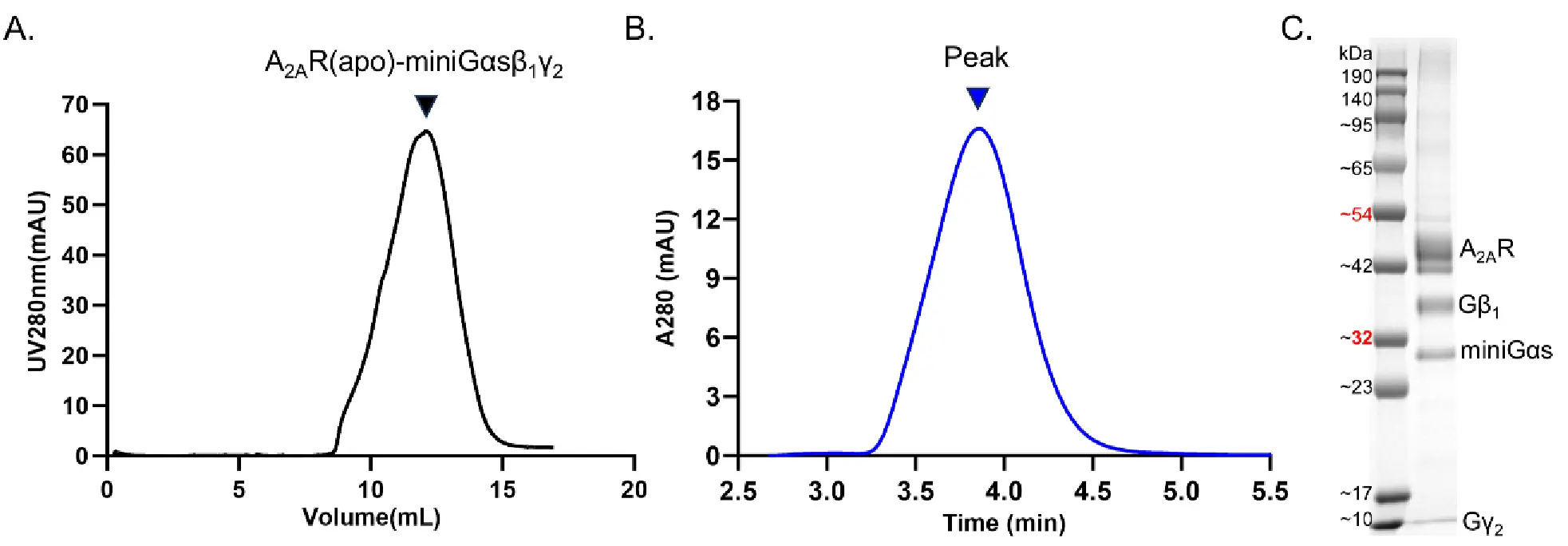
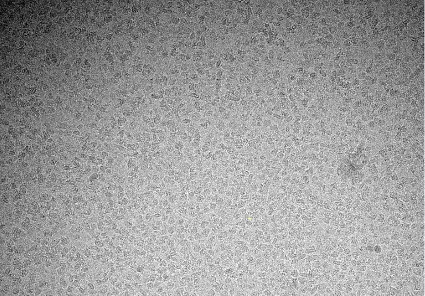
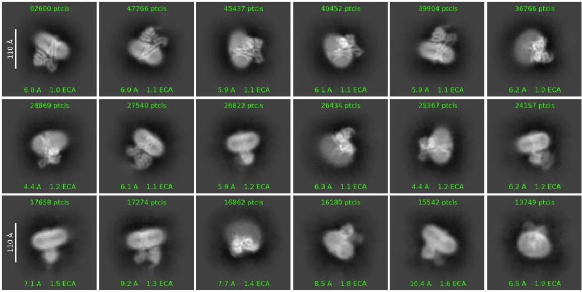
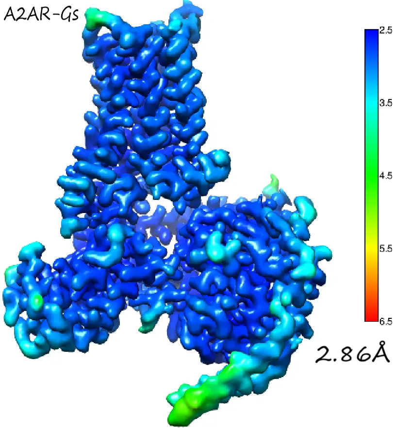
Introduction:
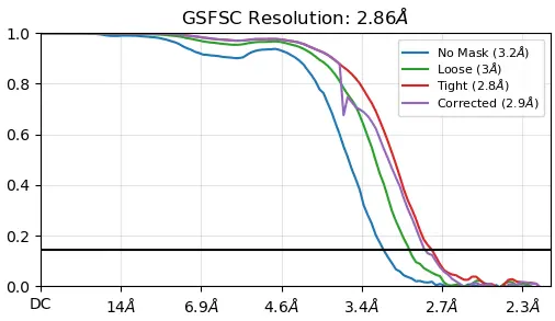
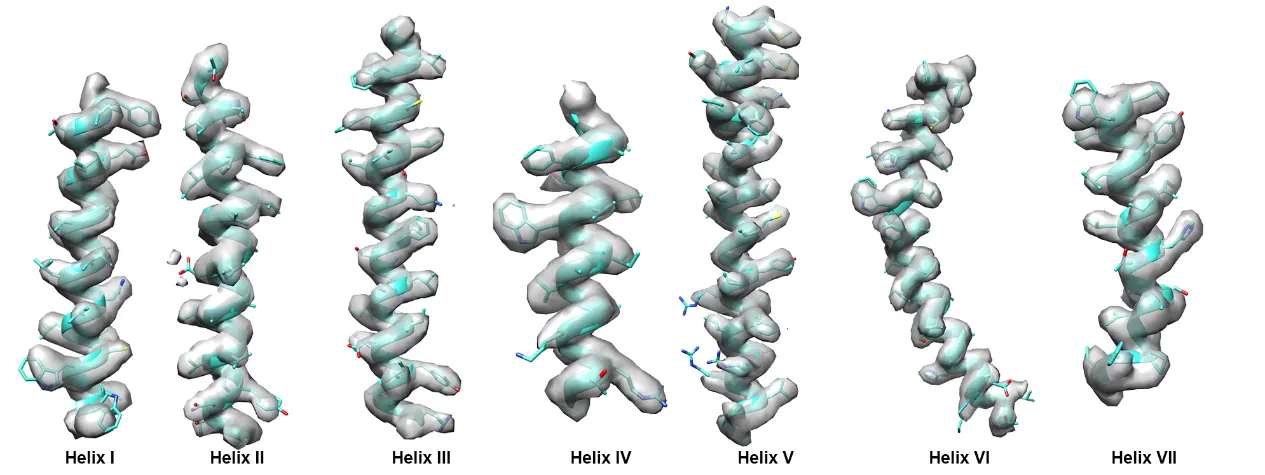
Distolife membrane protein R&D team purified the target membrane protein—the A_2AR−Gscomplex—using a detergent and phospholipid system. Through cryo-EM structural determination, they solved the 3D protein structure at a resolution of 2.86 Å. This structure shows high consistency with previously published literature while achieving superior resolution.
Case 2: Cryo-EM Structure of GCGR-Gs Complex
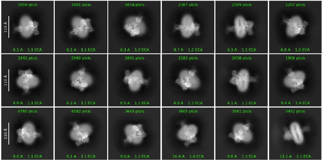
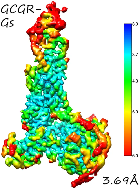
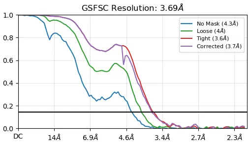
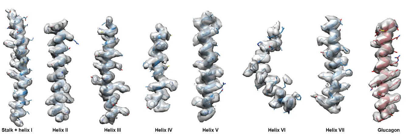
Introduction:
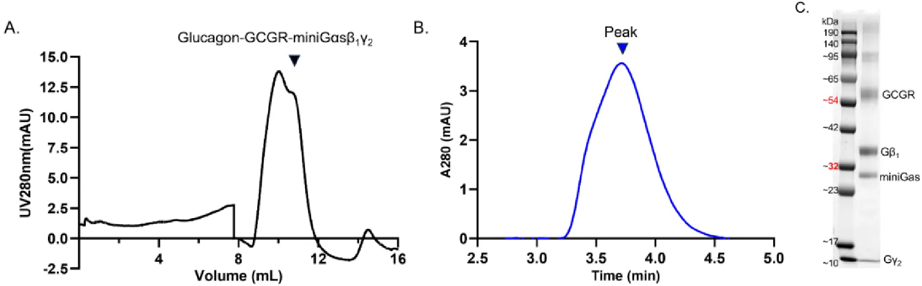
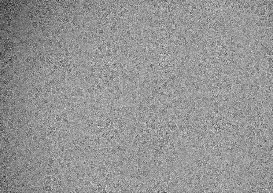
Distolife membrane protein R&D team purified the target membrane protein—the GCGR-Gs complex—using the LMNG and CHS system. Through cryo-EM structural determination, they resolved the 3D protein structure at a resolution of 3.69 Å. This structure shows high consistency with previously published literature while achieving superior resolution with a smaller dataset.
Case 3: Cryo-EM Structural Analysis of GCGR-Gs and GCGR-Gi1 Complexes
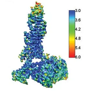
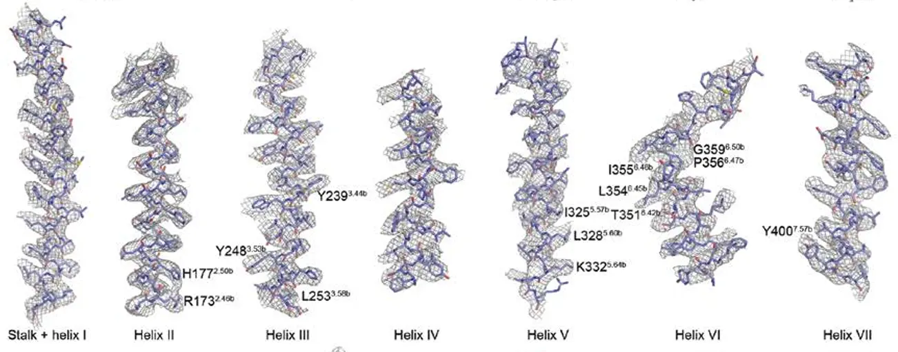
Introduction:
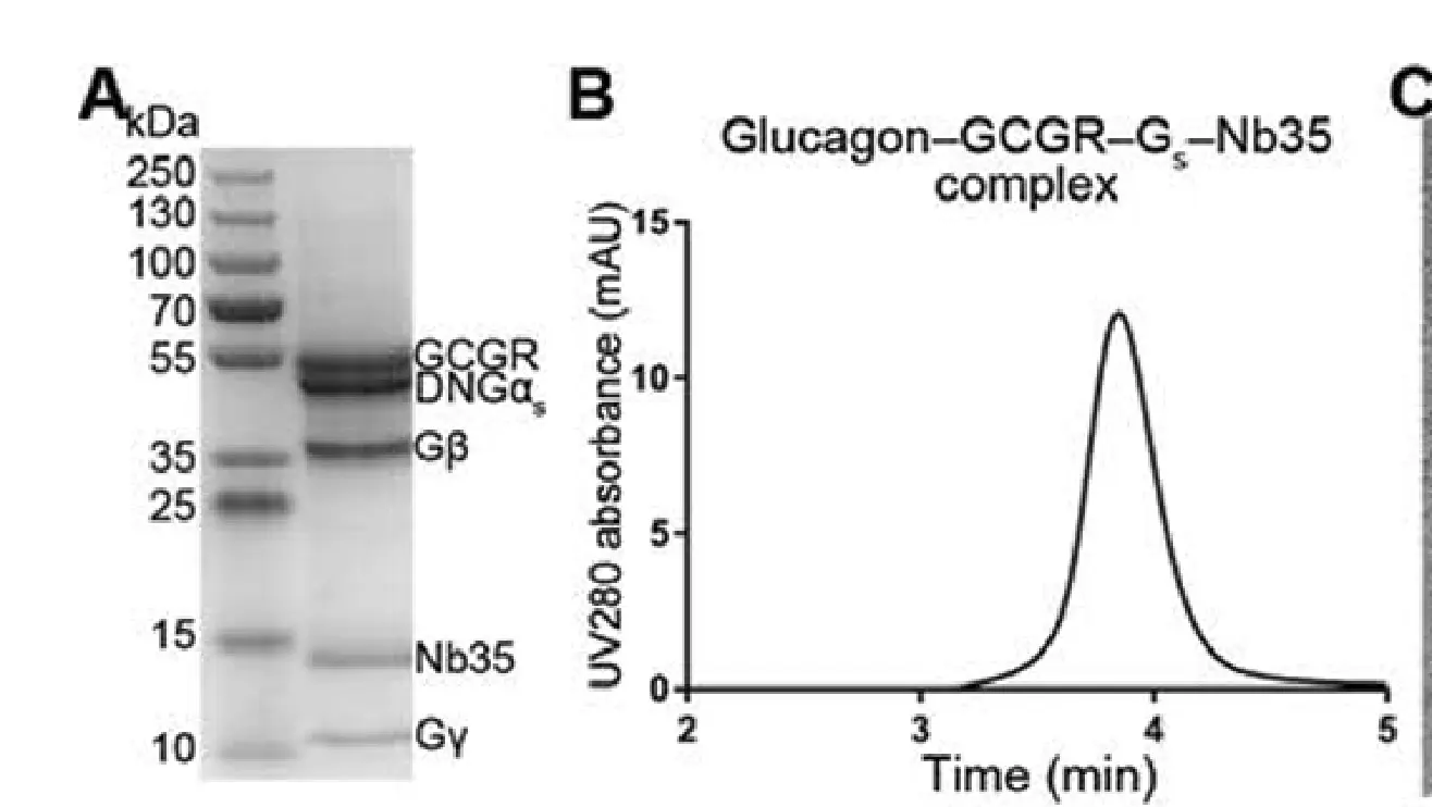
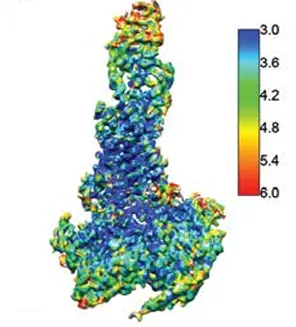
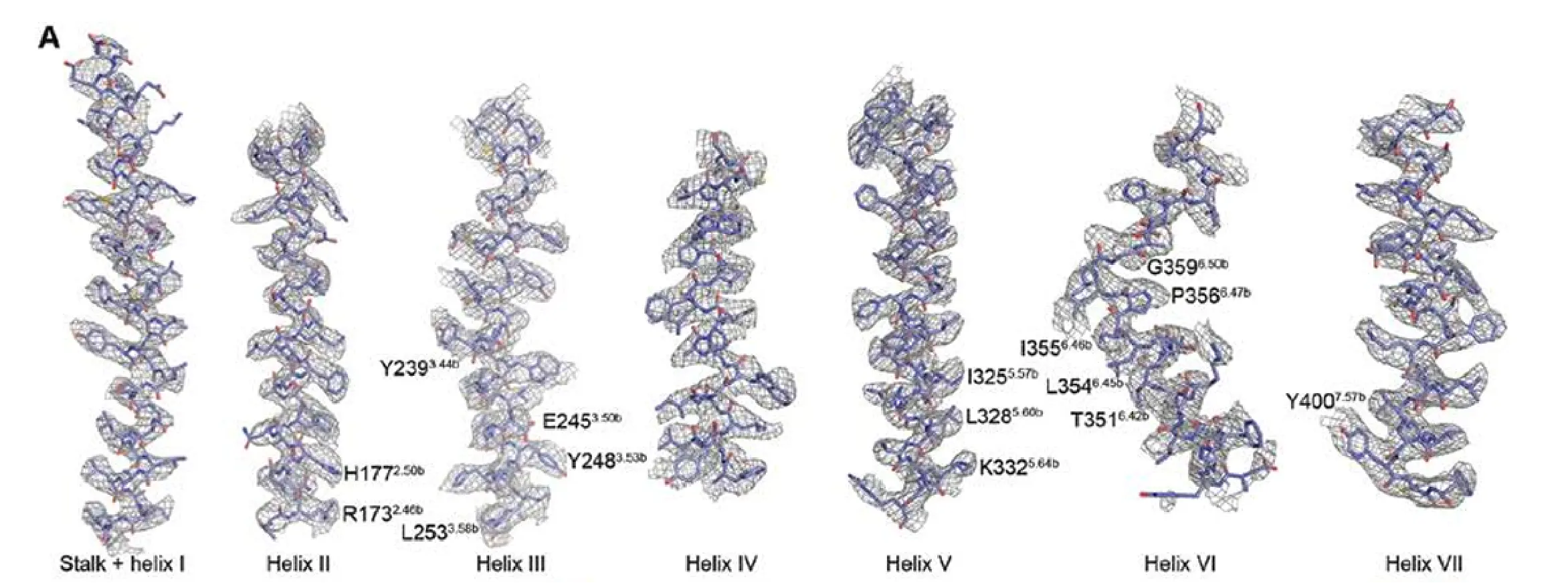
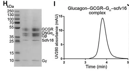
The Chief Scientific Officer of the Distolife team used cryo-EM to resolve, for the first time, the complex structures of a Class B GPCR—the human glucagon receptor (GCGR)—bound to Gs and Gi1, respectively (3.7/3.9 Å). Combined with NanoBiT, mutation, and signaling assays, the study revealed that the receptor accommodates both G proteins using a common open pocket: Gs achieves biased coupling through a larger interface (802 vs 551 Ų), whereas differences in intracellular loops ICL1/ICL2 dictate selectivity. This revises the traditional model that "pocket size determines specificity," laying a structural foundation for biased drug design.
Case 4: Cryo-EM Structural Analysis of mGlu2–Gi1 and mGlu4–Gi3 Complexes
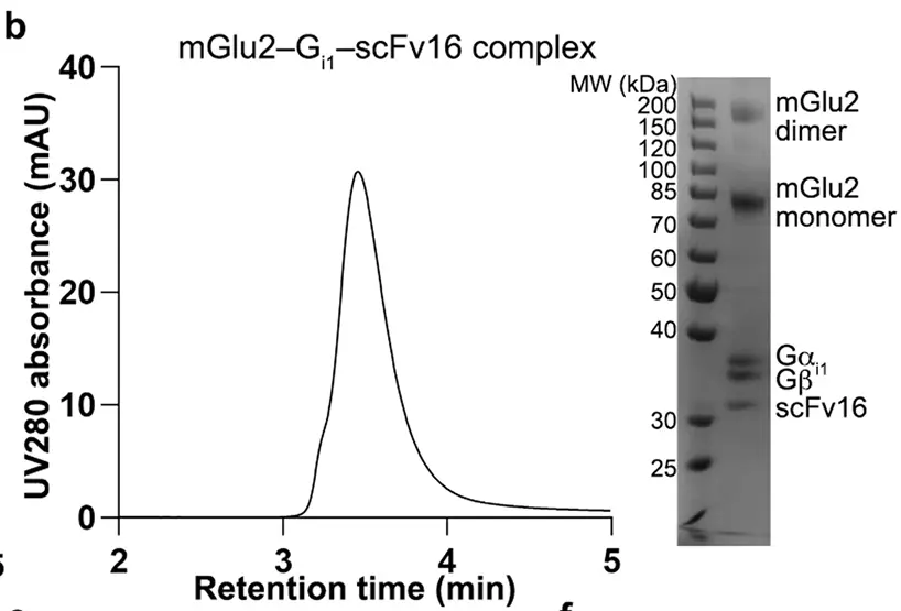
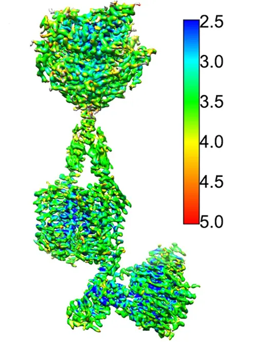
Introduction:
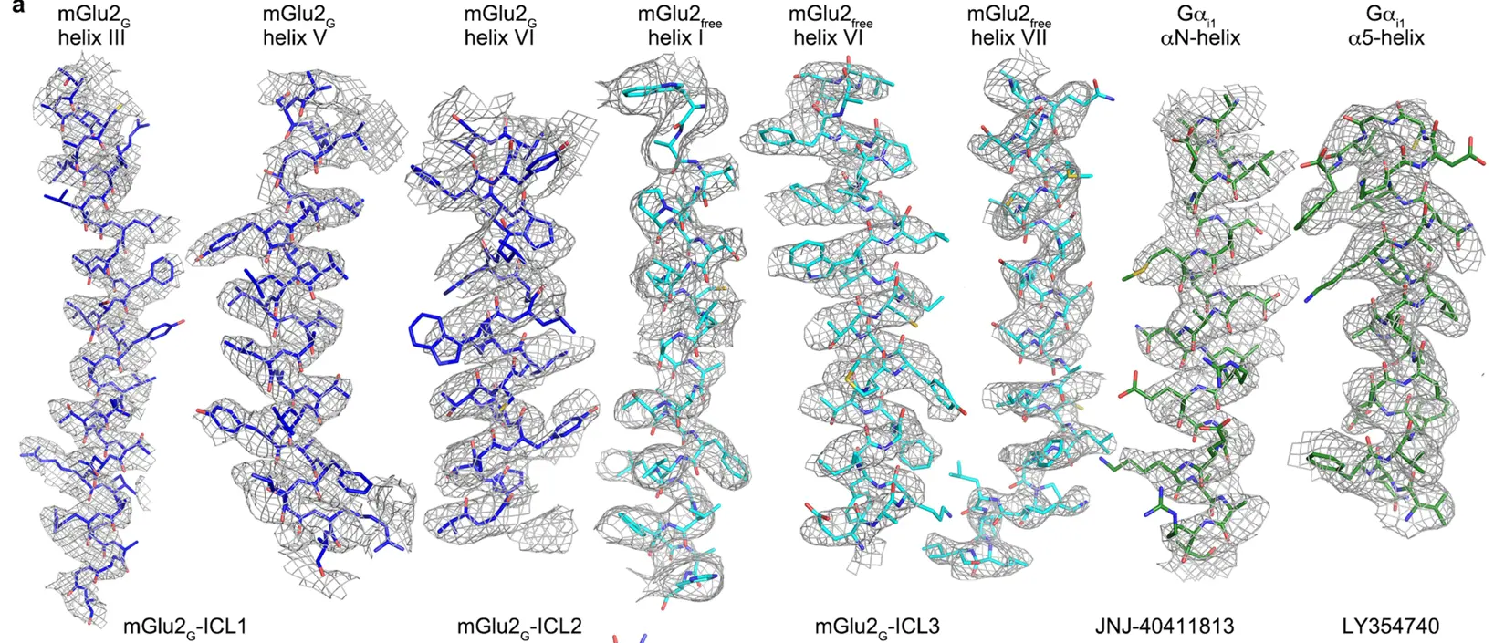
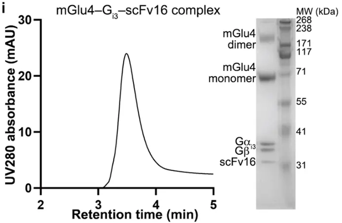
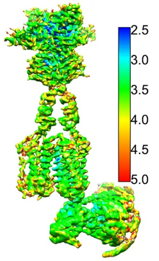
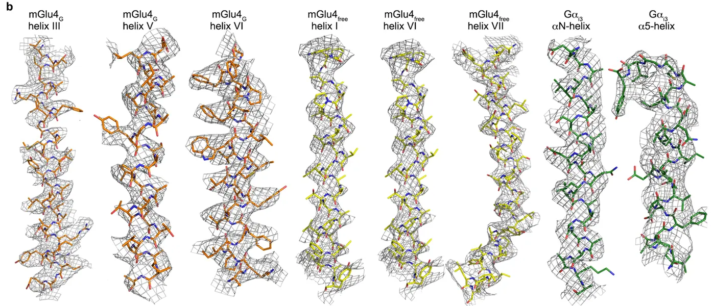
The Chief Scientific Officer of the Distolife team used cryo-EM to resolve, for the first time, the fully active complex structures of Class C GPCRs—human mGlu2 and mGlu4 bound to Gi proteins (3.1−4.0Å. Combined with mutagenesis, NanoBiT, and cysteine cross-linking assays, the study revealed that TM6 does not shift outward upon mGlu activation, and the α5C-terminus of Gαibinds to a shallow groove formed by three intracellular loops together with helix III/IV. Furthermore, it demonstrated at atomic resolution for the first time that the asymmetric dimer interface serves as the structural basis for "only one receptor subunit coupling to a G protein," deepening the understanding of GPCR activation.
Case 5: Crystal Structure Determination of Y1R in Complex with Selective Antagonists
Introduction:
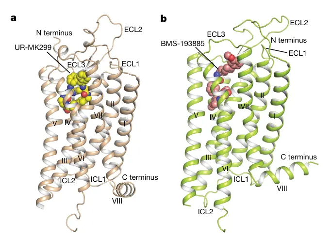
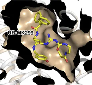
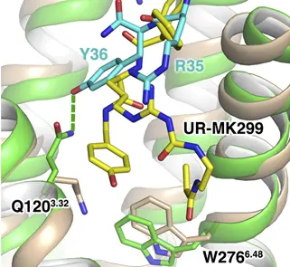
The Chief Scientific Officer of the Distolife team determined the first crystal structures of the neuropeptide Y receptor family—human Y1R in complex with two selective antagonists (2.7/3.0 Å.). Combined with mutagenesis, solid-state NMR, Rosetta docking, and photo-crosslinking, the study elucidated the antagonist binding modes and key selectivity determinants, such as the D287^6.59salt bridge. Furthermore, it identified for the first time the interactions between the N-terminus of the endogenous agonist NPY and the receptor's N-terminus as well as ECL2, laying a structural foundation for designing NPY-targeted drugs against obesity and other diseases.
Leave a Message
Inquiry Submitted
Thank you for your inquiry. Our team will get back to you as soon as possible.